CBCT in Vienna: 3D Diagnostics and Radiation Protection
When a 3D image genuinely informs the decision — and when two-dimensional imaging is enough.
10–40-second scan
Only the necessary field
Reduced-dose protocol
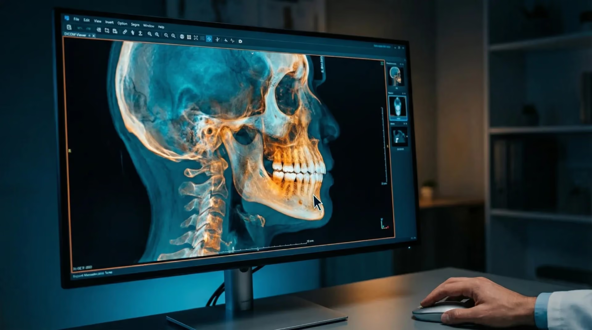
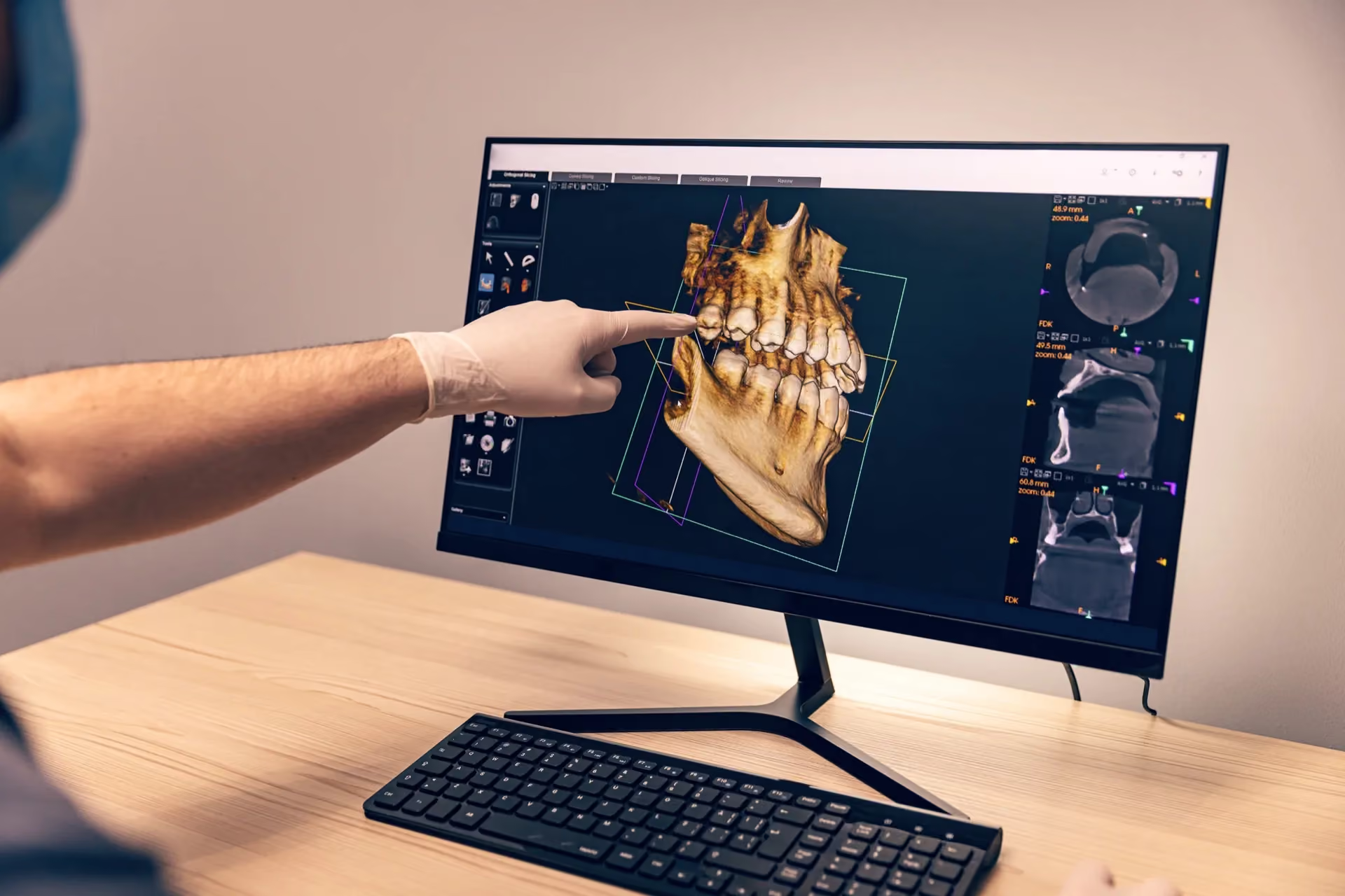
Discuss findings on screen
CBCT X-rays Vienna
dvt-roentgen-wien
4.9★
Doctify rating
25+
Years of experience
Personal
In-practice consultation
1010 Vienna
Kärntner Straße 9
At a glance
What is CBCT?
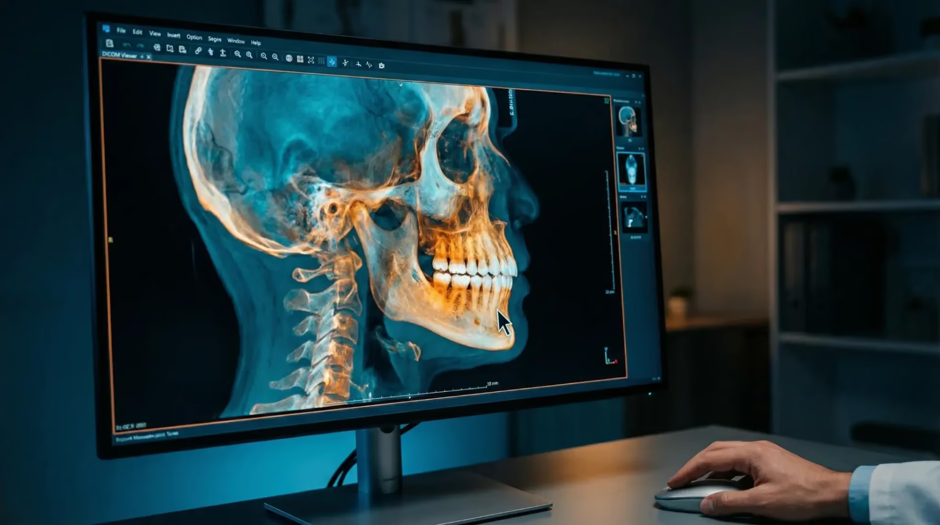
Cone-beam computed tomography is a three-dimensional X-ray method used in dental, oral and jaw care. It captures a volume in one rotation and shows the spatial relationship between nerves, bone and roots.*
Is CBCT dangerous?
CBCT uses X-rays and therefore requires a specific reason. A review reported effective doses from 34 to 1073 microsieverts depending on equipment and settings; the imaging field is therefore kept as small as possible.*
How does CBCT differ from CT?
Both provide three-dimensional X-ray images. CBCT captures a volume using a cone-shaped beam, while CT calculates sectional images. The number of rotations does not establish a universal dose ranking. Suitability and dose depend on the question, device, field and protocol; another examination may be more appropriate for soft tissues.*
When is CBCT needed?
When a two-dimensional image cannot answer a spatial question, such as whether a wisdom tooth lies against a nerve. A meta-analysis of three studies reported sensitivity for predicting nerve exposure of 95.1% with CBCT and 73.9% with panoramic imaging. This does not prove prevention of nerve injuries.*
Seeing more does not automatically mean better treatment.
Information that supports a decision
A panoramic image compresses a three-dimensional jaw onto a flat view. What lies in front or behind cannot be distinguished. This can matter when assessing whether a root touches a nerve or lies two millimetres away.
Why we still take a cautious approach
Every image involves radiation, which needs justification. We identify the question first and check whether an existing image already answers it.

Cone-beam computed tomography
We assess whether a 3D image would genuinely help answer the question.
Further detail
What CBCT actually contributes
The value of CBCT is well documented, but not equally for every purpose. It depends on whether the clinical question requires spatial information.
A clear example is assessment before wisdom tooth removal. A meta-analysis compared the methods for predicting exposure of the lower jaw nerve during surgery: CBCT achieved sensitivity of 95.1 percent, compared with 73.9 percent for panoramic imaging (Reia et al., 2021). The analysis included three studies and concerned prediction of nerve exposure, not proof that CBCT prevents nerve injury. In implant planning, reduced-dose protocols provide measurements with a pooled error of −0.24 millimetres (Carneiro et al., 2024), relating to the linear bone measurements studied. Bone strength or quality cannot be determined from these measurements alone; examination and overall planning also matter.
An honest account of radiation dose
There is no single CBCT dose. The literature reports a range of 34 to 1073 microsieverts for dental scans (Baena-de la Iglesia et al., 2022), depending on device, field size and protocol. This means dose can be adjusted. A systematic review of 16 studies found similar results for several objective outcomes with reduced-dose protocols, although subjectively assessed image quality could be worse (Kaaber et al., 2024). The key question is whether device, field and protocol answer the clinical question with sufficient image quality. These results establish neither general equivalence between all protocols nor routine follow-up CBCT without a separate indication.
The image alone does not decide
Two recent studies highlight the role of the clinician interpreting the images. Eight readers assessed the same 30 panoramic and 30 CBCT images; oral and maxillofacial surgeons identified nerve canal displacement more frequently than general dentists (Couto et al., 2025). A University of Zurich research model was trained on 4516 images to identify signs on two-dimensional images that might indicate three-dimensional imaging (Carvalho et al., 2023). It does not replace dental assessment of whether CBCT is necessary for the specific question. Both support the same principle: indication before equipment.
When CBCT may be considered
These treatments do not automatically require CBCT. It may be considered when a specific diagnostic question needs spatial information and existing images are insufficient, for example in implant planning, before wisdom tooth removal where nerve proximity is suspected, before root-end surgery, for complex root canals or assessment of a jaw cyst. Individual justification is assessed from the question; field size and protocol should fit. YERIT explains the method in more detail in its guide to 3D dental X-rays (CBCT).
YOUR CONCERNS
What is on your mind?

Information before surgery
A wisdom tooth may lie close to a nerve, or implant planning may need spatial bone measurements. CBCT can help clarify position; bone quality and strength are not inferred from these measurements alone.
A finding remains unclear
A two-dimensional image shows something that cannot be adequately assessed. We establish whether a 3D image would genuinely add information.
A second opinion using your image
You bring an existing image. We first check whether it answers our question; if so, no new image is needed.
SUITABILITY CHECK
Is this treatment right for you?
YOUR OPTIONS
TREATMENT OPTIONS COMPARED
We will discuss payment terms with you prior to treatment.
The indicative market prices in our price overview provide an initial guide. We will discuss which services are planned for you and the costs involved based on your examination findings and individual cost plan.
Advanced Dental Aesthetics & Implantology
Priv.Doz.Dr.Dr. K. Yerit
“Aesthetics is not a coincidence, but the result of precise planning and deep passion.”
CBCT is justified by a spatial clinical question, rather than by availability. If a two-dimensional image answers it, no additional scan is needed.
View profile →Scientific references
Institute for Implantology and Digital Dentistry Vienna
Medizinische Universität Wien
Österreichische Gesellschaft für Mund-, Kiefer- und Gesichtschirurgie (ÖGMKG)
European Academy of DentoMaxilloFacial Radiology (EADMFR)
Österreichischer Verband für Strahlenschutz
Yerit Practice – Dentist Vienna
Excellence
Why Smile4Life
Indication before equipment
We identify the question first. If a two-dimensional image answers it, we stop there.
Small field, appropriate low-dose protocol
Only the relevant area is imaged. Reduced-dose protocols may be appropriate if they answer the question with sufficient diagnostic quality. This must be checked for the device, field and protocol.
Specialist interpretation
Doz.Dr.Dr. Yerit interprets the scan as an oral and maxillofacial surgeon, considering the clinical question.
Frequently asked questions
Frequently asked questions
Do you have a specific question about your diagnosis? Book a consultation appointment directly →
Questions about your treatment?
Discuss what matters to you and what the next steps look like with us.
Book your appointment nowYour orientation
The scan itself takes 10 to 40 seconds; saved images may help later treatment. Before repeating imaging, we check whether suitable existing images answer the current question.
Understanding what’s involved
One appointment. Preparation, scanning, processing and discussion of findings take around 20 to 30 minutes together.
In everyday life
Who this information is for
For patients with a spatial question: a wisdom tooth’s proximity to the lower jaw nerve, bone height and width before an implant, cyst extent, complex or curved root canals, or unclear findings after dental injury. Also where an existing image does not answer the question.
Important limitations
During pregnancy, non-urgent imaging is postponed; if it cannot be delayed, justification is assessed particularly strictly. The same standard applies more strictly to children and adolescents because growing tissues are more sensitive. We do not scan if a single-tooth film or existing image already answers the question, or if the result would not change management.
Our approach
We regard CBCT as a useful tool for a limited range of questions. Where those questions apply, benefit is documented: sensitivity for predicting nerve exposure was 95.1 versus 73.9 percent compared with panoramic imaging (Reia et al., 2021). This measures prediction of exposure, not prevention of nerve injury. Without a relevant question, another image adds radiation rather than benefit.
We are equally open about dose. Rather than giving one reassuring-sounding number, we cite the literature’s range of 34 to 1073 microsieverts (Baena-de la Iglesia et al., 2022) and explain its basis: device, field and protocol. These must fit the question. Reduced-dose protocols can yield similar results for certain outcomes, while required diagnostic quality must be maintained (Kaaber et al., 2024). Each repeat image needs its own justification.
SCIENTIFIC STUDIES
We regard CBCT as a useful tool for a limited range of questions. Where those questions apply, benefit is documented: sensitivity for predicting nerve exposure was 95.1 versus 73.9 percent compared with panoramic imaging (Reia et al., 2021). This measures prediction of exposure, not prevention of nerve injury. Without a relevant question, another image adds radiation rather than benefit.
We are equally open about dose. Rather than giving one reassuring-sounding number, we cite the literature’s range of 34 to 1073 microsieverts (Baena-de la Iglesia et al., 2022) and explain its basis: device, field and protocol. These must fit the question. Reduced-dose protocols can yield similar results for certain outcomes, while required diagnostic quality must be maintained (Kaaber et al., 2024). Each repeat image needs its own justification.
Kaaber L et al. (2024). Low-dose CBCT protocols in implant dentistry: a systematic review
A review of 16 studies reports similar results for certain objective measurement tasks with reduced-dose and higher-resolution CBCT protocols. Subjective image quality could be worse. Whether a protocol adequately answers the specific diagnostic question must be assessed individually. The authors call for further clinical studies; follow-up CBCT also needs its own indication.
38679501
Carneiro ALE et al. (2024). Accuracy of linear measurements for implant planning based on low-dose cone beam CT protocols
The review included 13 studies, nine in the meta-analysis, with 81 specimens in total. For linear bone measurements using reduced-dose CBCT protocols in implant planning, pooled measurement error was −0.24 millimetres (95% confidence interval −0.52 to 0.04), tending towards underestimation. Exceptions occurred in the posterior mandible. Linear dimensions alone establish neither bone quality nor load-bearing capacity; applicability depends on the question and protocol (Dentomaxillofacial Radiology, 2024).
38429951
Reia VCB et al. (2021). Diagnostic accuracy of CBCT compared to panoramic radiography in predicting IAN exposure
A meta-analysis compared the methods for predicting exposure of the lower jaw nerve during wisdom tooth removal. Three-dimensional imaging achieved sensitivity of 95.1%, compared with 73.9% for panoramic imaging. Specificity was 64.4% versus 24.8% (Clinical Oral Investigations, 2021).
34275000
Baena-de la Iglesia T et al. (2022). Diagnostic performance of cone-beam computed tomography to diagnose root resorption
A systematic review with meta-analysis of 17 papers found sensitivity of 78.12% and specificity of 79.25% for detecting external root resorption. It reports effective doses for dental CBCT of 34 to 1073 microsieverts, depending on equipment, field size and protocol (Journal of Evidence-Based Dental Practice, 2022).
36914301
Couto DAF et al. (2025). Comparison of panoramic radiography and cone beam computed tomography for lower third molar assessment
Eight readers assessed 30 panoramic and 30 CBCT images from the same patients. Specialty measurably affected findings: oral and maxillofacial surgeons identified nerve canal displacement and narrowing more often than general dentists, while radiologists identified more root curvature (Journal of Oral and Maxillofacial Surgery, 2025).
40446846
Carvalho JS et al. (2023). Preinterventional third-molar assessment using robust machine learning
A model trained on 4516 panoramic images from the University of Zurich’s dental centre achieved accuracy of 0.94 and 0.93 for assessing nerve superimposition and root development respectively. The authors derive a table indicating when additional three-dimensional imaging may be useful (Journal of Dental Research, 2023).
37944556
Öffentliches Gesundheitsportal Österreichs: Digitale Volumentomographie (DVT)
Cone beam computed tomography produces three-dimensional X-ray images using a cone-shaped beam and is mainly used to answer specific spatial questions about teeth and bone. A single rotation does not establish a generally lower dose than CT: the device, field of view, protocol and required image quality are decisive. Another radiological examination may be more suitable for soft-tissue questions (Gesundheitsportal Österreich; assessment based on the S2k-Leitlinie DVT).

The question before the scan
A panoramic image shows the jaw in two dimensions. CBCT adds a spatial view when that information is needed for the specific clinical question.
More topics in this area
Deepen your knowledge – find all information regarding the treatment process, costs, and results.

Kärntner Straße 9
100 meters from Stephansplatz, Innere Stadt.
Partnerships & Memberships & References