Intraoral Scanner Vienna: Impressions Without Impression Material
What digital scanning can do, and where conventional impressions remain the better choice
Duration depends on scan scope
No impression tray
No radiation
Accuracy depends on application
Intraoral Scanner Vienna
zahnscanner
4.9★
Doctify rating
25+
Years of experience
Personal
In-practice consultation
1010 Vienna
Kärntner Straße 9
At a glance
What is an intraoral scanner, and how long does scanning take?
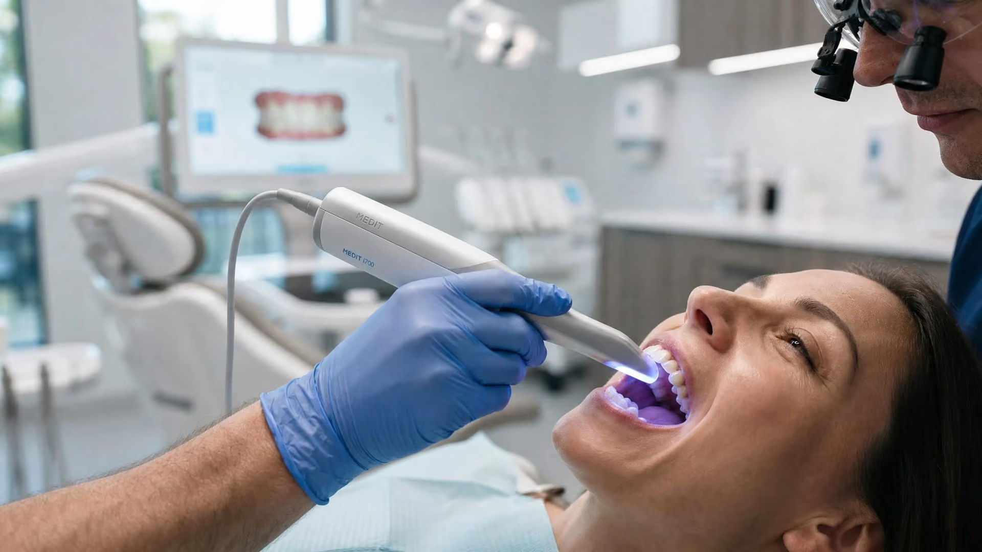
It records teeth and gums using light and a camera and calculates a digital 3D model. It uses no X-rays and can replace a conventional impression. Duration depends on recording individual teeth or both jaws and any rescanning. An overview of ten reviews found average time savings compared with impressions that must set in the mouth.*
What happens at a scan appointment?
You sit in the dental chair as usual. Teeth are dried, then the handpiece passes over chewing and side surfaces tooth by tooth; the bite is recorded last. You watch the model develop on screen, with missing areas rescanned. Scanning can be paused if you need a break.
Does a dental scan involve radiation?
No. An intraoral scanner records surfaces with light and a camera, without X-rays. Do not confuse it with CBCT, a three-dimensional X-ray method: both are sometimes called scans but are different examinations.*
From scan to finished restoration
How the data are used
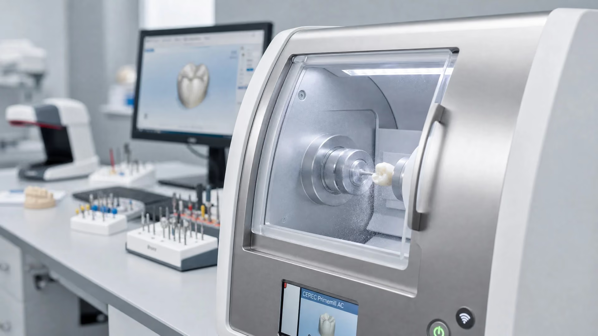
Scans are taken for crowns, inlays and bridges, aligners and retainers, bite splints and implant restorations. Data then go to the in-house milling unit or dental laboratory, depending on the restoration.
An underestimated benefit
Scans can be compared. Images from different times may document tooth-position and gum-surface changes. Meaningful comparison requires suitable superimposition and assessment alongside clinical findings.

Impression material
The 0.09 millimetres is an average study value comparing digital and alginate complete-arch impressions. The review includes 22 in-vivo studies and, in its clinical interpretation, concerns a fully natural dentition. The value is not an accuracy guarantee for every scan or implant case.
Basics
What is an intraoral scanner?
An intraoral scanner is a handheld optical device that records teeth and gums inside the mouth and calculates a digital 3D model. It replaces the tray and impression material for most treatments.
The handpiece projects a fine light pattern onto teeth and gums while recording how it deforms on the surface. Software calculates each point’s position repeatedly each second. Individual images are assembled continuously, like a phone panorama, while you watch the jaw model develop on screen. Intraoral means inside the mouth. For partial arches, the study found intraoral scanning more accurate than laboratory scanning of a model outside the mouth (Shah et al., 2023).
Trueness and precision: why both matter
Manufacturers often shorten this to “accuracy”, but two concepts are involved. Trueness describes how close the result is to reality. Precision describes how similar repeated scans of the same situation are. A device can repeatedly make the same error: high precision, poor trueness. An overview of ten systematic reviews therefore reports both measures separately (Singh et al., 2025).
Further detail
What evidence shows intraoral scanning can do
The question is rarely whether a scanner is good enough, but what it is good enough for. The answer varies considerably with the starting situation.
For fully dentate natural arches, Kong’s review compared digital and conventional complete-arch impressions. It included 22 in-vivo studies, with 13 in the meta-analysis. Tooth widths and arch distances showed no significant difference; mean three-dimensional deviation versus alginate was 0.09 millimetres, and both methods achieved precision below 0.1 millimetres (Kong et al., 2022). These findings concern natural dentitions and alginate, not every prepared crown margin, implant restoration or impression material. Whether a scan is sufficient for your crown, inlay or aligners depends on the specific restoration and scan quality.
Two limits we discuss openly
For an edentulous jaw, the picture changes. Scans remain comparable at clearly identifiable hard-tissue structures, but substantial deviations occur at functional borders, the internal seal and soft palate (Srivastava et al., 2023). A scanner records only what it can see, and mobile mucosa is difficult to capture.
For a complete implant-supported arch, a systematic review included 13 studies: ten laboratory and three clinical. Stereophotogrammetry was more accurate in ten studies. Some tested intraoral scanners exceeded an angular deviation of one degree discussed there; this does not establish a general in-house manufacturing limit or promise a passive fit (Pozzi et al., 2025). Splinted scan bodies measurably improve results (Shetty et al., 2025), but a try-in remains sensible.
What changes in everyday treatment?
An overview of ten systematic reviews mainly reported shorter treatment times and greater comfort than conventional impressions. Included work varied in quality and frequently relied on laboratory studies; this does not establish a fixed duration or benefit for every application (Singh et al., 2025). Another practical advantage is targeted rescanning of missing or inadequately captured areas. Whether an additional scan, new recording or another impression is needed depends on quality and the planned restoration. The YERIT intraoral scanner guide explains the technology in detail.
YOUR CONCERNS
What is on your mind?

Gagging during impressions
Conventional impressions feel difficult. Scanning needs no tray or setting time and can be paused.
Tooth replacement is planned
A crown, inlay or bridge is planned. The scan provides the basis and may permit single-visit manufacture in suitable cases.
Monitor changes
Recession or tooth movement needs observation. Suitably superimposed scans can document changes and complement clinical examination.
SUITABILITY CHECK
Is this treatment right for you?
YOUR OPTIONS
TREATMENT OPTIONS COMPARED
We will discuss payment terms with you prior to treatment.
The indicative market prices in our price overview provide an initial guide. We will discuss which services are planned for you and the costs involved based on your examination findings and individual cost plan.
Advanced Dental Aesthetics & Implantology
Priv.Doz.Dr.Dr. K. Yerit
“Aesthetics is not a coincidence, but the result of precise planning and deep passion.”
Patients feel the comfort advantage. The clinical advantage is different: a scan can be repeated without putting an impression tray in the mouth again.
View profile →Scientific references
Institute for Implantology and Digital Dentistry Vienna
Medizinische Universität Wien
Österreichische Gesellschaft für Zahn-, Mund- und Kieferheilkunde (ÖGZMK)
Digital Dentistry Society (DDS)
European Association for Osseointegration (EAO)
Yerit Practice – Dentist Vienna
Excellence
Why Smile4Life
Studied accuracy
For natural arches, the review reported mean deviation of 0.09 millimetres versus alginate. It included 22 in-vivo studies, with 13 in the meta-analysis; this does not promise accuracy for every scan.
Openly discussed limits
We explain limitations for edentulous jaws and full implant arches and choose another method when appropriate.
Repeatable without another impression
Incomplete areas can be rescanned if needed. Whether to add or repeat a scan depends on quality and planned restoration.
Frequently asked questions
Frequently asked questions
Do you have a specific question about your diagnosis? Book a consultation appointment directly →
Questions about your treatment?
Discuss what matters to you and what the next steps look like with us.
Book your appointment nowYour orientation
Duration depends on recording scope and extra scans. Stored data may support later comparisons, whose value depends on suitable superimposition and clinical findings.
Understanding what’s involved
One appointment, usually within already planned treatment. No preparation or after-effects.
In everyday life
Who this information is for
For treatments needing a model of a dentate jaw: crowns, bridges, inlays, onlays, veneers, aligners, retainers, bite splints and single-tooth implant restorations. Particularly useful for pronounced gag reflex or fear of conventional impressions, and for monitoring over years.
Important limitations
For complete dentures in edentulous jaws, conventional functional impressions are often better because scanners cannot reliably capture functional borders. Additional methods are used for full arches on several implants. Heavy gum bleeding, deeply subgingival margins and severely limited mouth opening complicate scanning; we treat the cause first or choose another method.
Our approach
We use intraoral scanning routinely where evidence supports it: for natural arches, a review of 22 in-vivo studies, including 13 in the meta-analysis, reported mean deviation of 0.09 millimetres versus alginate and precision below 0.1 millimetres for both methods (Kong et al., 2022). This is a study average for that setting. Suitability for your restoration is assessed separately; scanning can provide impressions without a tray.
We also explain its limits. In edentulous jaws, it fails where mobile mucosa needs capturing (Srivastava et al., 2023). For full implant arches, stereophotogrammetry was more accurate in ten of 13 studies, ten laboratory and three clinical (Pozzi et al., 2025). Conventional impressions or another method may then be preferable, and we choose accordingly even if less modern in appearance.
SCIENTIFIC STUDIES
We use intraoral scanning routinely where evidence supports it: for natural arches, a review of 22 in-vivo studies, including 13 in the meta-analysis, reported mean deviation of 0.09 millimetres versus alginate and precision below 0.1 millimetres for both methods (Kong et al., 2022). This is a study average for that setting. Suitability for your restoration is assessed separately; scanning can provide impressions without a tray.
We also explain its limits. In edentulous jaws, it fails where mobile mucosa needs capturing (Srivastava et al., 2023). For full implant arches, stereophotogrammetry was more accurate in ten of 13 studies, ten laboratory and three clinical (Pozzi et al., 2025). Conventional impressions or another method may then be preferable, and we choose accordingly even if less modern in appearance.
Kong L et al. (2022). Digital versus conventional full-arch impressions in linear and 3D accuracy: a systematic review and meta-analysis of in vivo studies
The review compared digital and alginate full-arch impressions of natural dental arches. It included 22 in-vivo studies, with 13 in the meta-analysis. Tooth widths and arch distances showed no significant difference; mean three-dimensional deviation was 0.09 millimetres, and precision for both methods was below 0.1 millimetres. These findings do not provide a general accuracy promise for prepared crown margins, implant restorations or other impression materials (Clinical Oral Investigations, 2022).
35786783
Singh R et al. (2025). Accuracy and clinical performance of intraoral scanners compared to conventional and extraoral impressions: an umbrella review
The review of ten systematic reviews from 2020 to 2024, involving over 30 device models, mainly reported shorter treatment times and greater comfort than conventional impressions. Many underlying investigations were laboratory studies, and the quality of the included reviews varied. Accuracy remains a limitation in toothless and partially dentate jaws. The findings guarantee neither a fixed duration nor freedom from discomfort for every scan (Cureus, 2025).
41141051
Pozzi A et al. (2025). Photogrammetry versus intraoral scanning in complete-arch digital implant impression
The review compared intraoral scanners and stereophotogrammetry for complete-arch impressions involving multiple implants. It includes 13 studies: ten laboratory and three in-vivo studies. Photogrammetry was more accurate in ten studies. The results and reported one-degree threshold are not a personal accuracy guarantee; further clinical studies and a try-in remain important.
40481748
Shah N et al. (2023). Validation of digital impressions accuracy obtained using intraoral and extraoral scanners: a systematic review
A systematic review of ten studies found that both scanner types fall within a clinically acceptable range. For partial-arch recording, the intraoral scanner is more accurate than an extraoral model scanner. Robust patient-based comparative data are lacking for the complete arch (Journal of Clinical Medicine, 2023).
37762774
Srivastava G et al. (2023). Accuracy of intraoral scanner for recording completely edentulous arches: a systematic review
A systematic review of twelve studies examined scanning of toothless jaws. Where hard-tissue structures were clearly identifiable, the scanner achieved accuracy comparable to conventional impressions. Marked deviations occurred at the functional border, internal seal and soft palate (Dentistry Journal, 2023).
37886926
Shetty PS et al. (2025). Accuracy of the digital implant impression with splinted and non-splinted intraoral scan bodies: a systematic review
A systematic review found that splinting scan bodies improves the accuracy of digital implant impressions, particularly for a complete arch. It stabilises the reference points and makes it easier for the scanner to stitch individual images together. However, some individual studies found no advantage (Journal of Indian Prosthodontic Society, 2025).
40654117
Öffentliches Gesundheitsportal Österreichs: Digitale Volumentomographie (DVT)
Cone beam computed tomography produces three-dimensional X-ray images using a cone-shaped beam and is mainly used to answer specific spatial questions about teeth and bone. A single rotation does not establish a generally lower dose than CT: the device, field of view, protocol and required image quality are decisive. Another radiological examination may be more suitable for soft-tissue questions (Gesundheitsportal Österreich; assessment based on the S2k-Leitlinie DVT).

A scan is the beginning, not the result.
Scan data are used to make crowns, inlays, aligners, retainers and bite splints, milled in-house or made in the laboratory. The scan is their common starting point.
More topics in this area
Deepen your knowledge – find all information regarding the treatment process, costs, and results.

Kärntner Straße 9
100 meters from Stephansplatz, Innere Stadt.
Partnerships & Memberships & References