Jaw Cysts in Vienna: Diagnosis and Treatment
Jaw cysts are assessed from your clinical findings and appropriate imaging. We discuss which treatment makes sense for you.
Imaging based on findings
Tissue always examined
Usually local anaesthesia
Follow-up by cyst type
Jaw Cyst Vienna
kieferzyste-wien
600
1200
4.9★
Doctify rating
25+
Years of experience
Personal
In-practice consultation
1010 Vienna
Kärntner Straße 9
At a glance
What is a jaw cyst and how serious is it?
A jaw cyst is a fluid-filled cavity with its own tissue lining.
It is usually benign but may grow unnoticed and affect bone, adjacent teeth or nerves. Tissue examination establishes which cyst is present; type and individual findings determine treatment and follow-up.*
How much time off is needed after jaw cyst surgery?
There is no fixed duration. It depends on cyst size and location, anaesthesia and your work. After general anaesthesia, do not drive for 48 hours; the treating doctor confirms sick leave.*
How is a jaw cyst treated?
The cyst is usually removed surgically and its tissue examined. Very large keratocysts are often decompressed or opened into the mouth first so they shrink, followed by removal and regular X-ray reviews.*
Further detail
Jaw cysts: a cavity that may cause no symptoms
Jaw cysts are fluid-filled cavities with their own tissue lining. They grow slowly, often cause few symptoms and displace bone without being noticed. Diagnosis is therefore frequently incidental, well before possible surgery. YERIT explains how cyst treatment fits within the wider specialty in its guide to oral and jaw surgery.
They are often discovered on an image taken for another reason: before wisdom tooth removal, implant planning or a review. By then, a cyst may have grown substantially and destroyed bone that later needs bone grafting.
A dentigerous cyst develops around the crown of an unerupted tooth, such as an impacted wisdom tooth. An analysis of 377 patients with histologically confirmed developmental odontogenic cysts included 286 with dentigerous cysts and 85 with keratocysts (Szaraz et al., 2025). This selected population does not represent all jaw cysts. Distinguishing these types matters for your care and cannot be done from an image alone.
Jaw cyst surgery: from imaging to review
Assessment begins with examination and appropriate imaging. The steps needed depend on size, location, type and existing images.
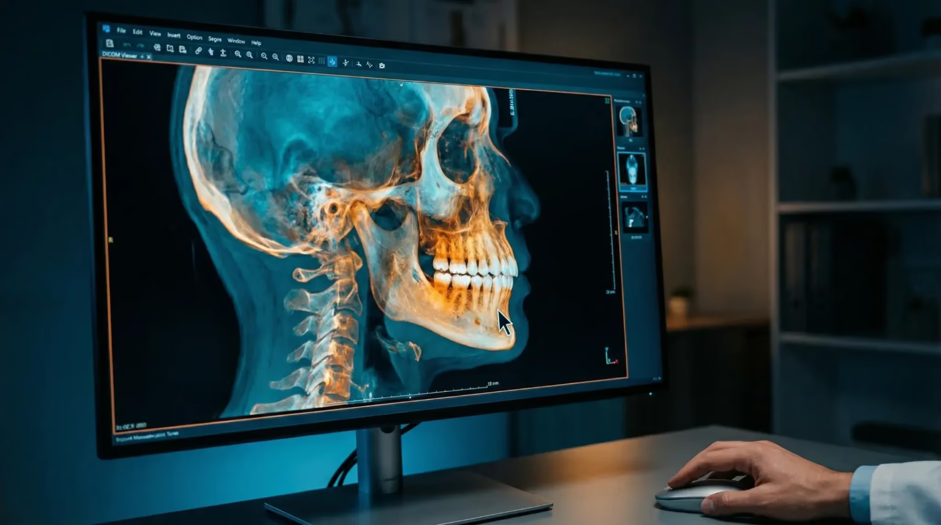
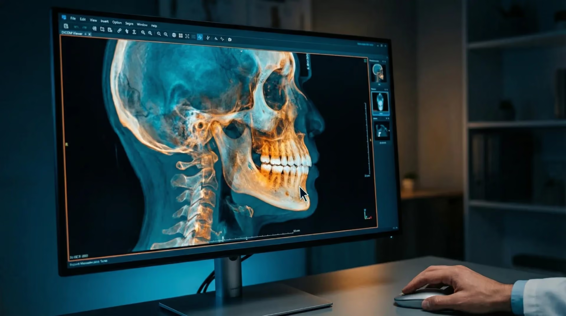
- Additional imaging: CBCT may be appropriate when existing images cannot adequately answer a specific clinical question about extent or position. It can then show the three-dimensional relationship to a nerve, sinus and adjacent roots.
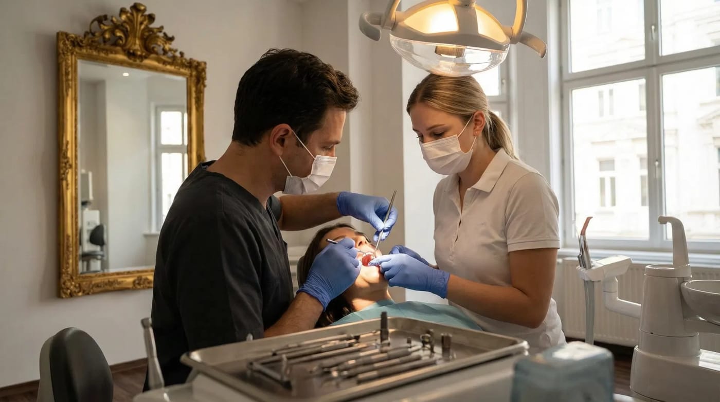
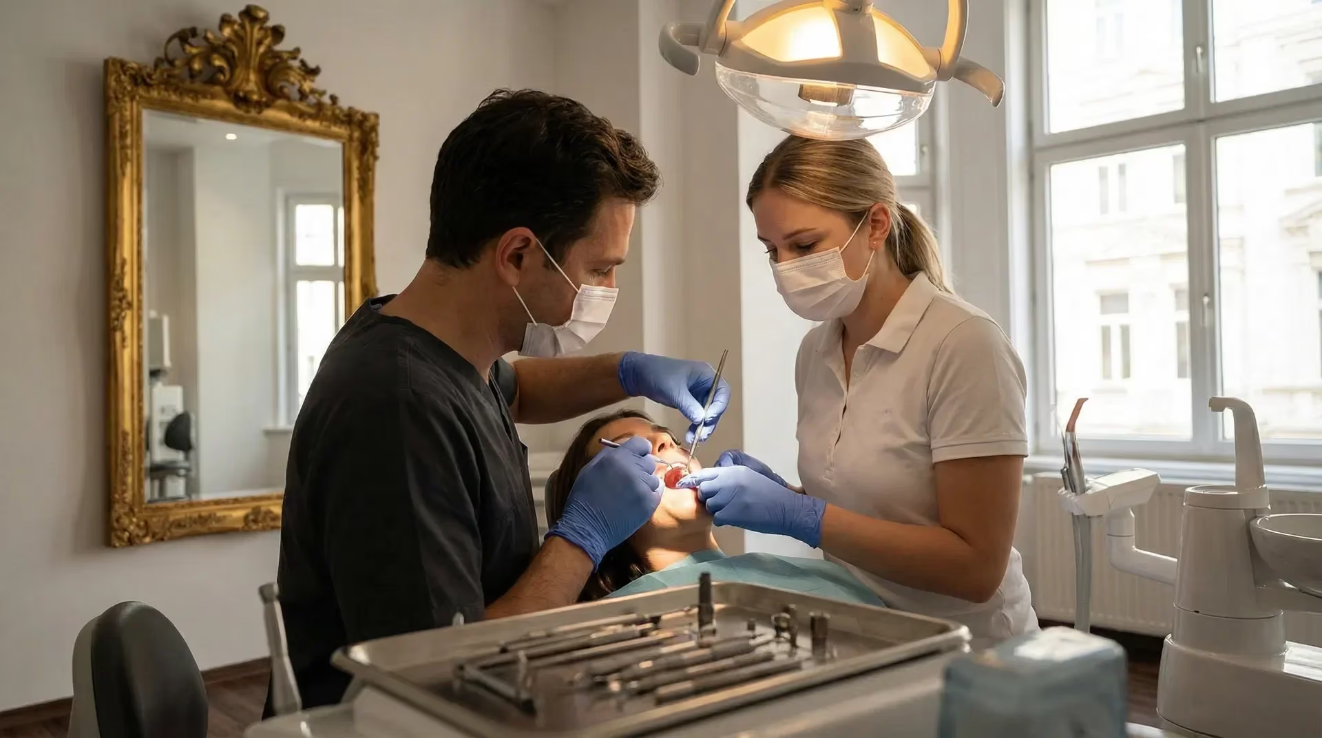
- Surgery: Usually under local anaesthesia, the cyst is completely removed (enucleated). Very large lesions are decompressed first and removed later.
- Laboratory findings: All removed tissue is sent to a pathology laboratory to identify cyst type.
- Reviews: The tissue result determines whether and how often X-ray reviews are needed; keratocysts require follow-up over several years.
The following sections explain why these steps matter.
When additional imaging may help
A panoramic image is a two-dimensional overview and may not adequately clarify the extent or spatial position of some lesions. Clinical examination, existing images and the specific question determine whether further imaging is needed and which method fits. Current ADA/AAOMR guidance recommends individual imaging selection for jaw lesions. When justified by a specific clinical question, CBCT can show the three-dimensional relationship to nerves, the sinus and adjacent roots (Benavides et al., 2026; PubMed).
For lesions in the back of the upper jaw, the relationship to the sinus may also matter. Additional imaging depends on examination, existing images and the unanswered question. See expertise for diagnostic technology.
What CBCT can show — and its limits
Three-dimensional imaging also helps characterise lesions. A study of 109 patients with lesions around impacted lower wisdom teeth found statistically significant differences between ameloblastoma, keratocyst and dentigerous cyst in location, internal density, bone expansion and direction of tooth displacement. Dentigerous cysts were limited to the crown in 88.9 percent, while all 14 ameloblastomas expanded the bone. The authors emphasise that several features must be considered together; one finding alone is insufficient for reliable classification (Li et al., 2023).
Even this combined assessment remains probabilistic. A meta-analysis of computer-assisted image interpretation estimated neural-network accuracy for distinguishing keratocyst from ameloblastoma on panoramic images at 0.893 — high, but not perfect (Fedato Tobias et al., 2024). Imaging alone therefore leaves diagnostic uncertainty.
Surgery: removal or decompression first
Small to medium-sized cysts are generally completely removed (enucleated) under local anaesthesia; the causative tooth is removed or root-treated. For very large lesions close to a nerve or replacing substantial bone, a two-stage approach may be gentler: first reducing the cyst through decompression, then removing it. A systematic review found lower recurrence with decompression without additional measures than with marsupialisation; where subsequent enucleation was performed, outcomes were similar (Tabrizi et al., 2019).
The main aim of staging is preservation: less bone loss, reduced risk to the lower jaw nerve and better prospects for adjacent teeth. Multiple keratocysts raise another consideration: in the analysis of 377 cases, 66 percent of patients with multiple keratocysts had Gorlin–Goltz syndrome (Szaraz et al., 2025). Assessment then extends beyond the jaw.
The laboratory provides the final classification
Every cyst specimen we remove is sent to pathology, even if imaging appeared clear. The lining is examined microscopically and classified under the WHO system. This result determines whether surgery completes treatment or whether years of follow-up are necessary.
The distinction is substantial: dentigerous and radicular cysts rarely recur after complete removal. For keratocysts, an overview of 19 systematic reviews reported a mean recurrence rate of 16.2 percent, rising to 43.2 percent after simple enucleation without additional measures (Gonçalves et al., 2024). Ameloblastoma requires a different assessment again. Tissue examination establishes which condition applies.
Costs and further planning
Non-binding Vienna market guidance for cyst surgery is around €600–1,200, depending on difficulty and whether bone grafting is also needed; the same range appears in our prices overview. Your individual plan specifies additional imaging, tissue examination and any further measures, including their charges. A binding figure requires examination because it depends on your findings; market ranges provide guidance only. We discuss your personal cost plan before surgery.
If an area of radiolucency has been found incidentally in your jaw, bring the image to your consultation and an older one for comparison if possible. Whether it has grown provides important information.
After surgery, follow the usual advice: cooling, soft food, no sport and no smoking for about a week. We discuss the laboratory result personally when available; it determines whether and how often follow-up is needed.

Wisdom teeth — each assessed individually
Not everyone has all four, and not every wisdom tooth needs removal. We assess each tooth individually according to its position, proximity to the lower jaw nerve and clinical findings.
YOUR OPTIONS
TREATMENT OPTIONS COMPARED
We will discuss payment terms with you prior to treatment.
The indicative market prices in our price overview provide an initial guide. We will discuss which services are planned for you and the costs involved based on your examination findings and individual cost plan.
Advanced Dental Aesthetics & Implantology
Priv.Doz.Dr.Dr. K. Yerit
“Aesthetics is not a coincidence, but the result of precise planning and deep passion.”
An image may show a cystic lesion, but it cannot establish exactly what it is. That is why every specimen I remove goes to the laboratory.
View profile →Scientific references
Institute for Implantology and Digital Dentistry Vienna
Medizinische Universität Wien
Österreichische Gesellschaft für Mund-, Kiefer- und Gesichtschirurgie (ÖGMKG)
European Association for Cranio Maxillo Facial Surgery (EACMFS)
WHO Classification of Head and Neck Tumours (5th edition, 2022)
Yerit Practice – Dentist Vienna
Frequently asked questions
Frequently asked questions
Do you have a specific question about your diagnosis? Book a consultation appointment directly →
Questions about your treatment?
Discuss what matters to you and what the next steps look like with us.
Book your appointment nowYour orientation
Understanding what’s involved
In everyday life
If an area of radiolucency has been found incidentally in your jaw, bring the image to your consultation and an older one for comparison if possible. Whether it has grown provides important information.
After surgery, follow the usual advice: cooling, soft food, no sport and no smoking for about a week. We discuss the laboratory result personally when available; it determines whether and how often follow-up is needed.
Who this information is for
For patients with a cystic jaw finding on an X-ray, often around an impacted wisdom tooth, at the root tip of a non-vital tooth or incidentally before implant planning. Also for people seeking another opinion on an existing finding.
Important limitations
Acute cyst inflammation is treated before enucleation. Very large lesions close to the lower jaw nerve are deliberately decompressed first rather than removed in one operation to protect the nerve. Blood thinners, bisphosphonate or antibody treatment for bone loss, and previous jaw radiotherapy require special planning and coordination with your treating doctor. Never stop blood-thinning medication yourself.
Our approach
SCIENTIFIC STUDIES
Szaraz D et al. (2025). Developmental odontogenic cysts with special focus on the occurrence of multiple cysts and syndromic association: a single-centre cross-sectional study from the Czech Republic
This cross-sectional university oral surgery study evaluates 377 patients with histologically confirmed developmental odontogenic cysts, all reclassified under the fifth WHO classification of 2022. Reported distribution: 286 dentigerous cysts, 85 keratocysts, 5 orthokeratinising cysts and one each of botryoid and calcifying cyst. For follow-up, 66 percent of patients with multiple keratocysts had Gorlin–Goltz syndrome, and recurrence was significantly associated with that syndrome. Multiple keratocysts therefore warrant additional specialist assessment (Orphanet Journal of Rare Diseases, 2025).
40038706
Li M et al. (2023). Cone-beam CT imaging features of common cystic lesions associated with the impacted mandibular third molar
This study of 109 patients at a university oral and maxillofacial radiology clinic compares CBCT features of three common lesions associated with impacted lower wisdom teeth: ameloblastoma, keratocyst and dentigerous cyst. Location, internal density, bone expansion and direction of tooth displacement differed significantly. Dentigerous cysts remained limited to the crown in 88.9 percent, while all 14 ameloblastomas showed outward or inward expansion. Several features must be considered together for reliable classification; one finding is insufficient (Chinese Journal of Stomatology, 2023).
37659849
Fedato Tobias RS et al. (2024). Diagnostic capability of artificial intelligence tools for detecting and classifying odontogenic cysts and tumors: a systematic review and meta-analysis
This meta-analysis assesses computer-assisted distinction of jaw cysts and tumours on X-rays and CBCT. For the particularly challenging distinction between keratocyst and ameloblastoma on panoramic images, neural networks achieved accuracy of 0.893 (95% confidence interval 0.832 to 0.954), with low-certainty evidence. CBCT datasets produced higher values based on only four papers. These findings illustrate the difficulty of image-based distinction; they concern diagnostic support from images and do not establish a replacement for tissue examination (Oral Surgery, Oral Medicine, Oral Pathology and Oral Radiology, 2024).
38845306
Gonçalves TOF et al. (2024). Management and recurrence of the odontogenic keratocyst: an overview of systematic reviews
This overview of 19 systematic reviews summarises recurrence of keratocysts after different methods. Mean recurrence was 16.2 percent; the highest reported value, 43.2 percent, followed simple enucleation without additional measures. Additional procedures and decompression before enucleation reduced recurrence. The authors also highlight critically low methodological quality, so the figures provide guidance rather than certainty (Oral and Maxillofacial Surgery, 2024).
38954313
Tabrizi R et al. (2019). Decompression or Marsupialization; Which Conservative Treatment is Associated with Low Recurrence Rate in Keratocystic Odontogenic Tumors? A Systematic Review
This systematic review compares two conservative approaches for large keratocysts: marsupialisation, opening the cyst into the mouth, and decompression through a drainage device. Without additional measures, decompression showed lower recurrence (p = 0.001). With subsequent enucleation, the difference disappeared. In extensive lesions, reduction before complete removal can help preserve bone, nerves and adjacent teeth (Journal of Dentistry Shiraz, 2019).
31579687
Kaimal VG, Patil B (2023). Evaluation of Association between Maxillary Posterior Teeth Periapical Pathologies and Maxillary Sinus Mucosal Changes-A Cone-Beam Computed Tomography (CBCT) Study
The CBCT study included 106 upper posterior teeth with periapical lesions. Mucosal thickening of the maxillary sinus was present at 99 teeth, or 93.4 percent. This was associated with proximity and damage to the separating bone wall. The study does not compare imaging methods and establishes neither general CBCT superiority nor symptom frequency across all jaw cysts.
38549905
Wisdom tooth removal – NHS
After wisdom tooth removal, pain and swelling for up to two weeks are common and usually improve after one to two days; after more difficult removal or general anaesthesia, one to three days off work are often needed. Do not drive for 24 hours after a sedative injection or 48 hours after general anaesthesia (nhs.uk, accessed 03.11.2026).
Imaging before the incision
For lower wisdom teeth, examination and suitable imaging help assess the position, roots and relationship to the lower jaw nerve. Additional CBCT is not routinely needed. Whether complete removal or, in selected situations, coronectomy is appropriate is decided individually.
More topics in this area
Deepen your knowledge – find all information regarding the treatment process, costs, and results.

Kärntner Straße 9
100 meters from Stephansplatz, Innere Stadt.

Partnerships & Memberships & References