
Apicectomy in Vienna: Procedure and Aftercare
What happens before, during and after surgery? These steps help you prepare and clarify questions at your appointment in Vienna.
Assess tooth preservation
Understand the procedure
Know the alternatives
Discuss questions personally
Apicectomy Procedure Vienna
wurzelspitzenresektion-ablauf-wien
4.9★
Doctify rating
25+
Years of experience
Personal
In-practice consultation
1010 Vienna
Kärntner Straße 9
At a glance
How is an apicectomy performed?
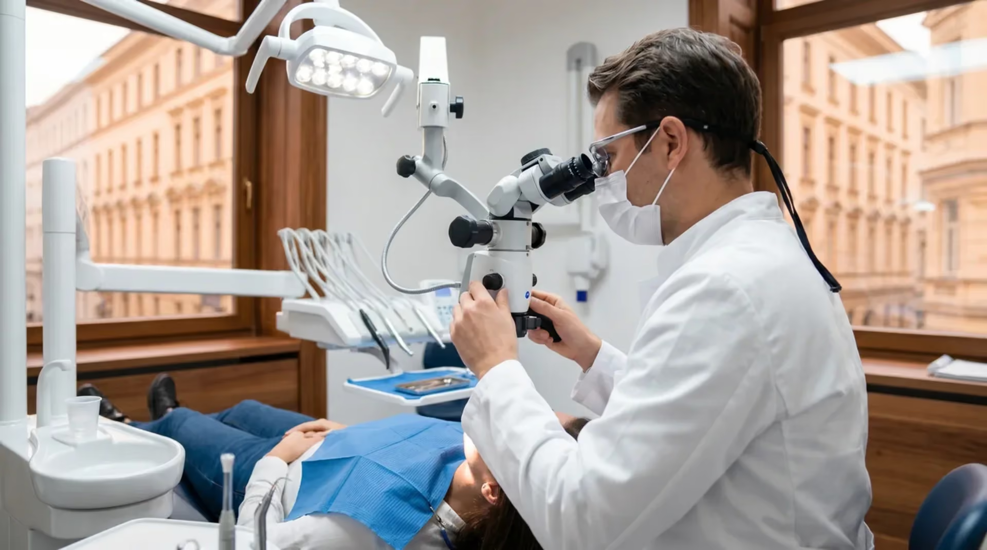
After examination and planning, the area is usually numbed with local anaesthesia. The root tip is reached through gum and bone. Inflamed tissue and a small part of the root tip are removed, and the canal end is sealed if needed. Wound closure and checks follow.
Wound pain and swelling usually subside after a few days. According to KZBV, stitches are removed after seven to ten days. The bone at the root tip takes considerably longer; an X-ray check after several months shows whether it is healing.
How long are you off work after an apicectomy?
There is no fixed duration. After an uncomplicated procedure under local anaesthesia, a short rest without physical exertion is often sufficient; heavy physical work or treatment of several teeth may require longer. Discuss the time you should pause after your procedure.
What should I prepare before the appointment?
Bring existing X-rays and findings, together with a current medication list. Be sure to discuss blood-thinning medication beforehand. Write down your questions, including about repeat root canal treatment as an alternative. Allow time to rest after surgery.
The procedure at a glance
From examination and choosing access through treatment of the root tip to wound healing, checks and subsequent restoration.

Apicectomy procedure: assessment and planning before surgery
The procedure begins by asking whether surgery is appropriate. Your dentist examines the affected tooth, assesses the existing filling or crown and previous X-rays. Additional imaging may be needed if findings are unclear.
A direct comparison of both approaches in 529 teeth has so far found no clear winner (Bucchi et al., 2022).
Discuss previous treatments, symptoms and your medication. Ask about alternatives too, particularly repeat root canal treatment. A list of your questions helps during the discussion. General information is available under apicectomy in Vienna. The YERIT guide Apicectomy procedure explains the individual steps in more detail.
How long does an apicectomy take?
This depends on tooth position, number of roots and intended access. A small randomised study of upper front teeth measured operating time: 39 to 47 minutes without a surgical guide and 26 to 27.5 minutes with one (El Marakby et al., 2026). These figures tell us little about molars or multiple roots.
[Source]Ask about the likely duration of your appointment and recovery afterwards. A time reported in a study cannot replace this planning.
Anaesthesia and access to the root tip
Apicectomy is usually carried out under local anaesthesia. The treated area should be largely pain-free. If you feel pain during treatment, tell the team immediately.
An incision in the gum provides access to the root area. The bone over the root tip is then opened carefully. The form of access depends, among other things, on the tooth’s position.
[Source]Removing the root tip and sealing the canal
Inflamed tissue and a small part of the root tip are removed, often around two to three millimetres. This describes the procedure generally, not a fixed amount for every tooth.
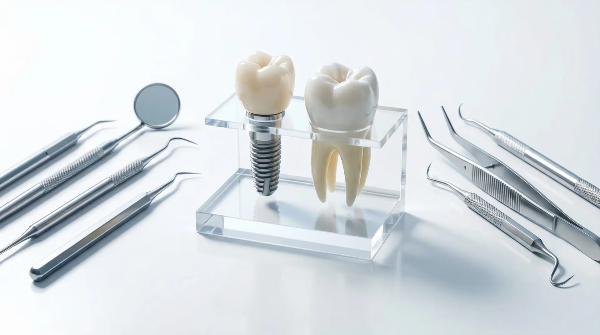
The root canal can then be prepared from its tip and sealed with a suitable material. This is called a retrograde filling: access is from the root tip. A review found no established superiority of the newer bioceramics studied over MTA.
[Source]A randomised study with 221 teeth at follow-up compared iRoot BP Plus with and without additional iRoot SP sealer. At twelve months, success rates were 94.5 and 92.8 percent respectively; the difference was not statistically significant (Dong et al., 2024). This does not establish identical prospects for every tooth.
An additional regenerative procedure may be considered for certain bone defects. Studies suggest a benefit, but do not mean that bone grafting is generally required. Whether such an addition is needed is decided separately.
[Source]Wound closure and healing
After cleaning and stopping bleeding, the gum is stitched. Gum and bone heal at different rates. KZBV gives seven to ten days until possible stitch removal and an X-ray check after three to six months as general guidance. Your appointments may differ.
[Source]A closed wound does not mean that inflammation in the bone has completely healed. Checks therefore remain important even when the tooth no longer hurts. Further treatment may be necessary if symptoms or abnormal findings persist. Healing also depends on the size of the original inflammation: lesions up to 5 mm heal markedly more often than larger ones in studies (Sabeti et al., 2023).
Aftercare: eating, oral hygiene and recovery
Wait until the anaesthetic wears off before eating. Soft food that is not hot is suitable initially. Cooling externally can ease symptoms. Continue cleaning your teeth carefully while protecting the operated area.
Smoking can interfere with wound healing. Avoid physical exertion initially and clarify when sport, heavy work and travel are appropriate again. Time away depends on your procedure and work; a fixed number of sick-leave days would be unreliable.
[Source]Take painkillers only according to the personal advice of your treating practice. A general article cannot determine which medicine suits your medication and medical conditions.
When should you contact the practice?
Swelling and discomfort can occur after surgery. If they become worse instead of better, the tooth throbs increasingly, pus appears or you develop fever, have the wound checked promptly. Persistent bleeding also needs dental assessment.
[Source] [Source]Possible risks also include wound-healing problems and injury to neighbouring structures. Depending on the tooth, proximity to the sinus or a nerve may matter. Your individual risks are discussed before surgery.
Discuss costs beforehand
The costs of apicectomy are clarified together with possible additional services. Non-binding average market guide prices are available on the price page. You receive your personal cost plan after examination.
Examination, possible surgery and follow-up checks are planned from your findings.
YOUR OPTIONS
TREATMENT OPTIONS COMPARED
We will discuss payment terms with you prior to treatment.
The indicative market prices in our price overview provide an initial guide. We will discuss which services are planned for you and the costs involved based on your examination findings and individual cost plan.
Advanced Dental Aesthetics & Implantology
Priv.Doz.Dr.Dr. K. Yerit
“Aesthetics is not a coincidence, but the result of precise planning and deep passion.”
The most important step happens before surgery.
View profile →Scientific references
Institute for Implantology and Digital Dentistry Vienna
Medizinische Universität Wien
Österreichische Gesellschaft für Mund-, Kiefer- und Gesichtschirurgie (ÖGMKG)
European Society of Endodontology (ESE)
Österreichische Gesellschaft für Endodontologie (ÖGEndo)
Yerit Practice – Dentist Vienna
Frequently asked questions
Frequently asked questions
Do you have a specific question about your diagnosis? Book a consultation appointment directly →
Questions about your treatment?
Discuss what matters to you and what the next steps look like with us.
Book your appointment nowYour orientation
Gain clarity before deciding.
Understanding what’s involved
The number of appointments depends on the findings and course of healing.
In everyday life
Contact your treating practice if symptoms increase, you develop fever or bleeding does not stop. Your personal aftercare instructions take priority over general advice.
Who this information is for
Important limitations
Our approach
SCIENTIFIC STUDIES
Ko MJ et al. (2025). Success rates comparison of endodontic microsurgery and single implants with comprehensive and explicit criteria: a systematic review and meta-analysis
The review combined separate study pools for endodontic microsurgery (22 studies) and single implants (6 studies). Using explicitly defined success criteria, it reported pooled microsurgery success of 89 percent, 90 percent with follow-up below five years and 80 percent from five years onwards; 78 percent was reported for single implants. These were not direct randomised comparisons or personal survival or cost forecasts. Suitable tooth-preserving or replacement treatment is chosen according to the clinical findings.
39979225
Sabeti M et al. (2023). The effect of hard tissue defects on the clinical outcome of endodontic microsurgery: a systematic review and meta-analysis
A meta-analysis of 19 studies estimated pooled success of endodontic microsurgery at 84.5 %. Lesion size was an important prognostic factor: lesions up to 5 mm healed in 78.4 % of cases, compared with 63.3 % for larger lesions (RR 1.12; p ≤ 0.05). Purely endodontic lesions tended to perform better than combined endodontic-periodontal lesions (81.4 % versus 68.2 %), but this difference was not statistically significant (Clinical Oral Investigations, 2023).
37932638
Bucchi C et al. (2022). Non-surgical root canal treatment and retreatment versus apical surgery in treating apical periodontitis: A systematic review
Five direct comparative studies involving 529 teeth and follow-up from six months to 8.7 years were assessed. Trends differed depending on the endpoint; neither option was clearly superior. Four studies had a high risk of bias and another raised concerns. Differences between studies prevented meta-analysis. This result does not establish equivalence for every clinical situation (International Endodontic Journal, online publication 2022, issue 2023).
35762859
Ibáñez-Aravena PC et al. (2025). New Bioceramics vs. Mineral Trioxide Aggregate (MTA) in the Success of Endodontic Microsurgery: A Systematic Review and Meta-Analysis
Five studies (three randomised controlled trials and two retrospective cohorts) compared newer bioceramics with established Mineral Trioxide Aggregate (MTA) for root-end filling. The meta-analysis found no significant difference in treatment success (RR 1.00; 95% confidence interval 0.94–1.07). Evidence quality was rated moderate. Newer bioceramics are therefore not demonstrably superior to MTA, but may be preferred because they are easier to handle (Australian Endodontic Journal, 2025).
40637350
Flynn R et al. (2024). The impact of bone grafting with/without barrier membrane placement on the outcome of apical surgery: A systematic review and meta-analysis
Eight randomised clinical trials assessed whether additional bone grafting — with or without a barrier membrane — improves healing after apicoectomy. The meta-analysis found a significant advantage for regenerative procedures over conventional surgery (odds ratio 2.18; 95% confidence interval 1.32–4.31; p = 0.004). Subgroup analyses did not establish an independent effect for individual bone substitute materials, membrane type or their combination (International Endodontic Journal, 2024). The paper does not justify routine use of these adjuncts in every apicoectomy; the authors call for further well-designed randomised trials.
38491954
Dong X et al. (2024). The outcome of combined use of iRoot BP Plus and iRoot SP for root-end filling in endodontic microsurgery: a randomized controlled trial
In this randomised controlled trial, 240 teeth with treatment-resistant inflammation at the root tip underwent microsurgery; 221 reached the twelve-month follow-up. Success rates in the two material groups were 94.5 % and 92.8 %, with no significant difference. Tooth type, a through-and-through bone lesion, the periodontal condition of the tooth and the Kim lesion classification were important factors affecting one-year success (Clinical Oral Investigations, 2024).
38430316
El Marakby MF et al. (2026). Assessment of guided versus non-guided endodontic microsurgery with root-end resection using different retrograde filling materials on operation time and lesion volume: A randomized controlled clinical study
In this randomised controlled study, 24 patients underwent apicectomy of upper front teeth with or without a surgical guide. Operating time measured during surgery was 39 to 47 minutes without a guide and 26 to 27.5 minutes with one. Bone lesion volume decreased markedly after six months in all groups without a between-group difference; the two filling materials performed comparably (Journal of Conservative Dentistry and Endodontics, 2026).
42004800
More topics in this area
Deepen your knowledge – find all information regarding the treatment process, costs, and results.


Kärntner Straße 9
100 meters from Stephansplatz, Innere Stadt.

Partnerships & Memberships & References